Metabolic responses in visual perception with fMRS
A collaborative research to advance our understanding of how the visual brain consumes energy to support neural function.
Makliya Mamat / June 8, 2024
What types of visual stimuli elicit the strongest responses in the brain? It’s well-documented that various visual stimuli can influence perception, but the specifics of which stimuli generate larger and more consistent responses have remained elusive.
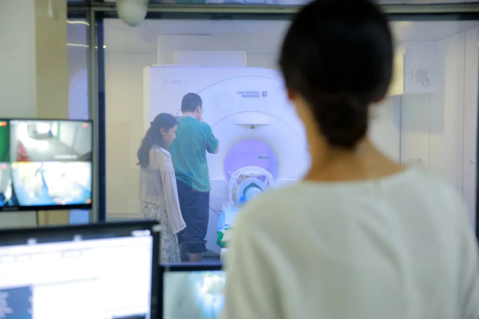
Recently, I had the opportunity to explore this question more deeply through a collaborative research project at Kangning Psychiatric Hospital. Working with researchers from Dr. Shen’s team and Zhiyong Zhang’s lab at Shanghai Jiao Tong University, the goal was to measure the metabolic changes in response to different types of visual stimuli and determine which ones produce larger and more consistent responses.

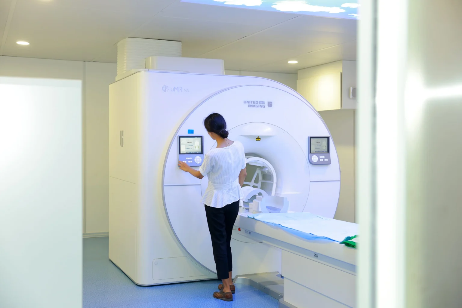
To tackle this, researchers can design various stimuli sessions for visual stimulation. Using functional magnetic resonance spectroscopy (fMRS) at higher field strengths like 5T, they could observe the metabolic changes that occur in response to these stimuli.
Unlike traditional fMRI, which focuses on blood flow changes, fMRS offers a window into the chemical environment of the brain, providing deeper insights into how different stimuli affect brain function at a metabolic level.
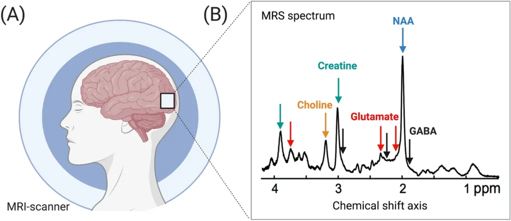
General scheme of MRS. (a) Metabolite signals are acquired using an MRS scan from a region of interest in the visual cortex (white box). (b) The MRS spectrum’s amplitude reflects the bulk signal across tissue types and cellular compartments. MRS Magnetic resonance spectroscopy, MRI magnetic resonance imaging, ppm parts per million. doi: 10.1007/s00429-021-02273-0
The preliminary experiments yielded some intriguing results. When comparing responses to non-sense images and “no-interesting” houses, faces consistently stimulated larger responses, with a dynamic increase in metabolic activity. This aligns with the theory that our brains are particularly attuned to recognizing and processing faces—a crucial ability for social interaction and survival throughout human evolution. The ability to measure these dynamic changes can lead to better diagnostic tools and therapies for conditions affecting visual perception and cognition.

One of the most rewarding aspects of this collaboration has been the interdisciplinary nature of the research team. Bringing together diverse perspectives and expertise, the synergy between clinical insights and advanced biomedical engineering techniques has been instrumental in driving the project forward.
Understanding the metabolic responses to different visual stimuli could pave the way for new approaches in treating psychiatric and neurological disorders. If we can identify specific visual stimuli that elicit beneficial responses, we could develop targeted therapies that leverage these stimuli to improve cognitive function and mental health.
Each new finding brings us closer to a more comprehensive understanding of the human brain and its remarkable capacity for perception and cognition. Reflecting on the resources and technology at our disposal, I am reminded of the scarcity of MRI machines and technicians in parts of the world like Africa. This makes me appreciate even more the resources we have and the importance of making the best use of them. It underscores the necessity of continued exploration and collaboration in neuroscience to ensure that these advancements reach as many people as possible.
Contents
Newsletter
- Updates from Makliya Notes will be delivered to your inbox.