“James Webb of the cell”
With its unique three-dimensional representation and analysis capabilities, this technology gives us a more real view of the cell world.
Makliya Mamat / December 8, 2022
“You are sitting at your microscope working at high magnification trying to sort out the three-dimensional compartmentalization of your fluorescent probe. You constantly focus up and down bringing different levels of the specimen into sharp focus while letting other levels above and below go out of focus. As this frustrating job continues, you say to yourself “It would be wonderful if I could see the whole specimen in clear focus all at once”. Confocal Laser Scanning Microscopy (CLSM) is the answer. ”
This beautiful passage from C. Robert Bagnell, Jr., Ph.D.’s introduction chapter for CLSM peaked my interest that I’m the one trying to create lab-based microscopy images with unparalleled depth and clarity. Well then, I had the good fortune to work with this incredible machine, which I like to refer to as the “James Webb of The Cell.”
Confocal microscopy technologies have been around for more than 40 years, but the growth and adaptability of the method have been fueled by improvements in optical design, spinning-disk technology, and camera technology.
In contrast to a wide-field microscope, where the entire specimen is illuminated by mercury or xenon and the image is visible to the naked eye, confocal microscopes use a different mechanism for creating images.
Illustrated in the figure below are a series of images that compare selected view-fields in traditional wide-field and laser scanning confocal fluorescence microscopy.
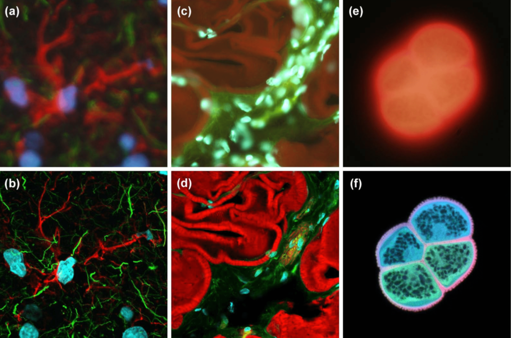
Comparison of widefield (upper row) and laser scanning confocal fluorescence microscopy images (lower row)
Researchers may create 3D images of organelles within living cells and study changes that take place in cells over time using confocal laser scanning and spinning-disk confocal microscopy. Multiple colors are used to show this information. In biology and the biomedical sciences, the method of spinning disk confocal fluorescence microscopy with laser scanning has become a crucial instrument.
Confocal microscopy has been widely used in medical and dental research and also for the clinical treatment of various diseases. Along with its research applications in cancer and Alzheimer’s disease.
CLSM is widely used in Europe and North America and is one of the most expensive types of microscope available in the market. However, it has not yet been used in scientific research in many regions of the world, in part due to its expense and in part due to lack of knowledge. Regardless of budgetary limitations, it is crucial that the local researcher have access to this amazing tool so that it can stake a position in contemporary literature.
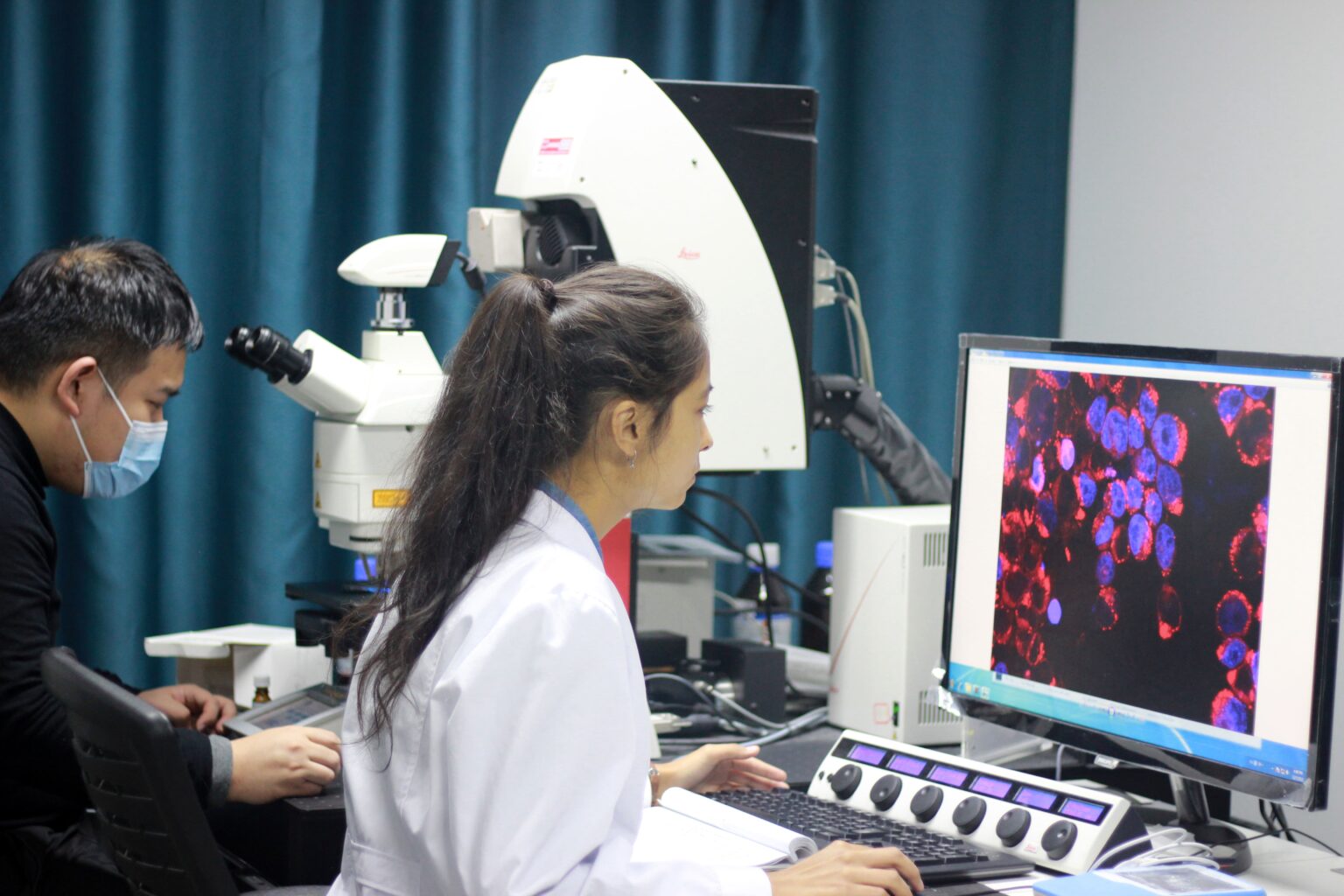
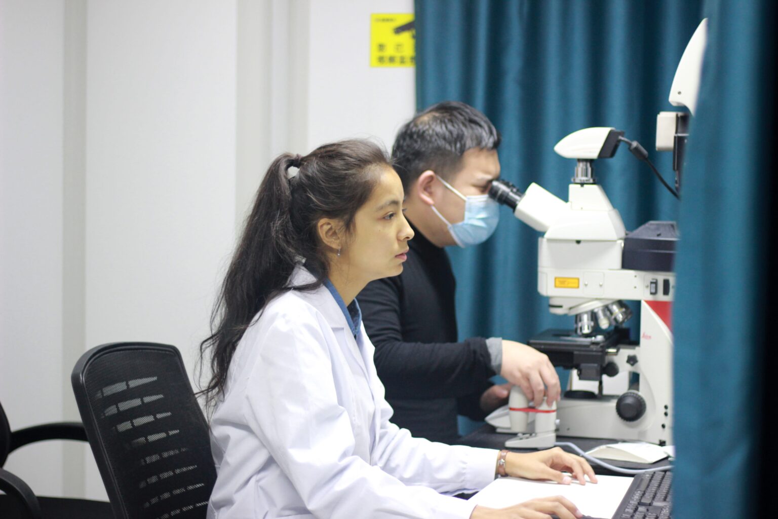
I’ve had the pleasure of working with Chen Wu. He is a kind individual who has persistently assisted me during our research by pointing out my mistakes (which I made a lot of) and shortcomings throughout hands-on experiments. I learned a lot from him, and enjoy the time spent in the laboratory.
Contents
Newsletter
- Updates from Makliya Notes will be delivered to your inbox.