A multi-omics approach
How mass spectrometry imaging through a multi-omics approach helps us better understand the brain’s complex molecular landscape.
Makliya Mamat / August 12, 2024
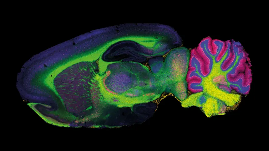
Imagine trying to understand a city by analyzing its power grid. You could look at the entire network at once or zoom in to see how individual homes and businesses are connected. Mass spectrometry imaging (MSI) works similarly, but instead of power lines, it maps the distribution of molecules within a biological sample at an extraordinarily fine spatial resolution.
At its core, mass spectrometry (MS) identifies and quantifies molecules based on their mass-to-charge ratio. MSI takes this a step further by adding spatial context, allowing us to create detailed maps of where specific molecules are located within a tissue, such as a section of the brain.
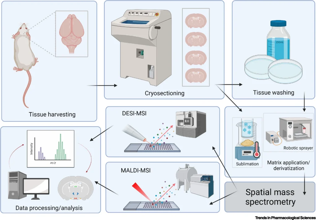
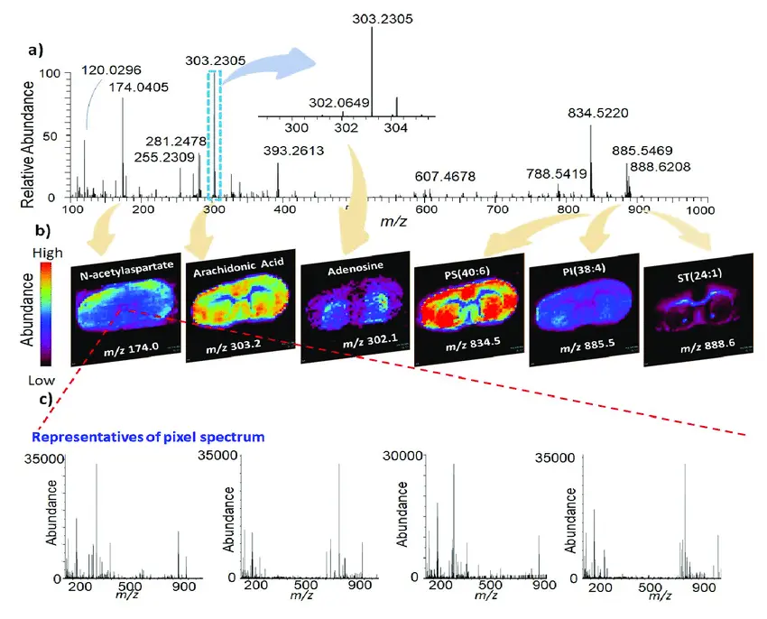
The process starts with preparing a thin tissue section, which is then placed in the mass spectrometer. A laser or ion beam bombards the tissue, ejecting molecules from the surface. These molecules are ionized and sent through a mass analyzer, where they are separated based on their mass-to-charge ratio. The result is a spectrum that reveals which molecules are present and, crucially, their precise location within the tissue.
Baijnath, Sooraj et al. Trends in pharmacological sciences vol. 43,9 (2022): 740-753.
Yan, Xin et al. Analytical chemistryvol. 92,19 (2020): 13281-13289.
Neuroimaging techniques like MRI offer a macroscopic view of the brain. Transcriptomics provides a snapshot of gene expression, giving us a molecular-level understanding of cell behavior.
MSI fills a crucial gap between these approaches by visualizing the biochemical landscape of the brain with spatial precision. For example, in neurodevelopmental disorders, where we know both cellular and molecular processes are altered, MSI can help identify specific lipid or metabolite changes that might drive these conditions.
If you have transcriptomics data that shows which genes are active in different brain regions. Layer on top of that MSI data showing where specific metabolites are concentrated. The result is a much richer, multidimensional map of the brain’s molecular landscape. This multi-omics approach is especially powerful in understanding complex conditions like Alzheimer’s disease, where changes occur simultaneously at multiple levels—genes, proteins, and metabolites.
The developments of integrating different “omics” approaches to build a comprehensive picture of brain function and dysfunction is promising. The brain is a black box in many ways, and tools like MSI with its ability to reveal unseen offer glimpses from different angles.
Search posts by date
Newsletter
- Updates from Makliya Notes will be delivered to your inbox.